Plasmapheresis for Optic Neuritis: Treatment and Recovery
5 min read
AUTHOR

MEDICALLY REVIEWED BY


Optic neuritis - inflammation of the optic nerve - is one of the most frightening neurological events a person can experience. Vision blurs, dims, or disappears, often accompanied by pain with eye movement. For most people, the first-line treatment is high-dose intravenous corticosteroids, which can accelerate recovery in milder cases. But when steroids fail to restore vision adequately, plasmapheresis for optic neuritis is increasingly considered as a second-line intervention.
This article explains what plasmapheresis involves, who the evidence suggests may benefit most, how plasma exchange for optic neuritis is typically performed, and what recovery can look like.
What Is Optic Neuritis?
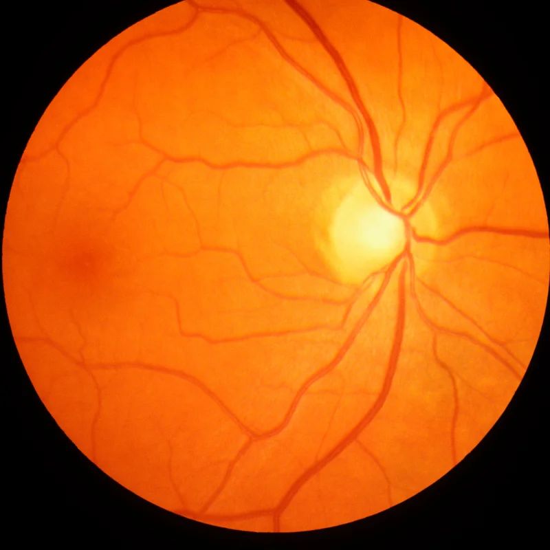
Optic neuritis is inflammation of the optic nerve, typically affecting one eye at a time. It causes a range of visual symptoms - from mild blurring to near-complete loss of central vision - along with pain behind the eye that worsens with movement. Color vision is often affected, with reds appearing washed out or dull.
The most common causes include:
- Multiple sclerosis (MS): Optic neuritis is the presenting symptom in roughly 15-20% of MS cases, and up to 50% of MS patients develop it at some point
- Neuromyelitis optica spectrum disorder (NMOSD): An antibody-mediated autoimmune disease associated with the AQP4-IgG antibody in approximately 73% of cases (Wingerchuk et al., Neurology, 2016)
- Myelin oligodendrocyte glycoprotein antibody disease (MOG-AD): A related but distinct condition also producing optic neuritis
In MS, optic neuritis often recovers spontaneously - though not always completely. In NMOSD, optic nerve attacks are typically more severe and are associated with a much higher rate of permanent visual damage if not treated aggressively. This distinction matters significantly when evaluating plasmapheresis.
How Plasmapheresis Works for Optic Neuritis
Plasmapheresis - more precisely called therapeutic plasma exchange (TPE) - removes a patient's plasma from the blood and replaces it with a substitute, typically human albumin. In doing so, TPE eliminates circulating proteins including autoantibodies, complement proteins, and inflammatory cytokines that may be driving or perpetuating the immune attack on the optic nerve.
The rationale for using plasma exchange for optic neuritis differs depending on the underlying diagnosis:
- In NMOSD, TPE directly removes the AQP4-IgG antibodies responsible for attacking astrocytes at the blood-brain and blood-spinal cord barriers. This makes the treatment mechanistically targeted - removing the pathogenic agent itself.
- In MS, the mechanism is less specific. TPE may remove complement, inflammatory mediators, and myelin-reactive antibodies, though the T-cell-mediated pathophysiology of MS is less directly addressed by plasma removal.
This biological difference helps explain why NMOSD-associated optic neuritis tends to respond more favorably to TPE than MS-associated optic neuritis. The Neuromyelitis Optica Study Group (NEMOS) recommends TPE as a first-line acute attack treatment in NMOSD, particularly for attacks that are severe or fail to respond adequately to IV methylprednisolone (Trebst et al., Journal of Neurology, 2014).
Why NMOSD Responds Differently Than MS
NMOSD is driven by a specific, identifiable antibody - AQP4-IgG (or MOG-IgG in seronegative cases) - that can be physically removed from the plasma. When TPE extracts this antibody, the immune attack on the optic nerve loses much of its driving force.
MS-associated optic neuritis, by contrast, is primarily T-cell mediated. While antibodies and complement play a role, removing plasma does not neutralize the core pathogenic mechanism as directly. The American Society for Apheresis (ASFA) reflects this in its guidelines: TPE is designated Category I or II for NMOSD attacks, and Category III (insufficient evidence; individualized decision) for MS-associated demyelinating disease, including optic neuritis (Connelly-Smith et al., Journal of Clinical Apheresis, 2023).
When Is Plasmapheresis Considered for Optic Neuritis?
Plasmapheresis for optic neuritis is generally considered in the following circumstances:
- After failure of high-dose IV methylprednisolone (typically 1,000 mg/day for 3-5 days)
- When vision loss is severe or functional vision is significantly compromised
- In NMOSD: may be initiated earlier in the attack course, sometimes concurrently with or shortly after steroids, given the severity of typical attacks
- When early intervention is possible: evidence suggests treatment within 20-30 days of symptom onset is associated with better visual outcomes (Borisow et al., Frontiers in Neurology, 2018)
Not all patients are candidates. TPE is contraindicated or requires careful evaluation in patients with active bleeding, hemodynamic instability, severe coagulopathy, or inability to tolerate central venous access. These are individualized decisions made by the treating neurologist in conjunction with the apheresis team.
What Does the Evidence Show?
The evidence base for plasmapheresis as a treatment for optic neuritis comes primarily from studies of CNS demyelinating disease broadly - MS and NMOSD - with optic neuritis as a common attack type.
The landmark randomized trial by Weinshenker et al. (Annals of Neurology, 1999) enrolled patients with severe, steroid-refractory acute CNS demyelinating attacks and found that 42% of patients receiving plasma exchange showed meaningful neurological improvement, compared to only 6% in the sham exchange group. Optic neuritis was among the attack types included.
Subsequent observational studies have extended these findings:
- Magaña et al. (Archives of Neurology, 2011) reported a 46.2% rate of functional improvement in a retrospective series of 59 patients with CNS demyelinating disease, including optic neuritis attacks.
- Llufriu et al. (Journal of Neurology, 2009) identified early treatment initiation and a favorable response after the first session as the strongest predictors of good outcome at 6 months.
- Kleiter et al. (Neurology: Neuroimmunology & Neuroinflammation, 2016) analyzed 207 apheresis interventions in NMOSD specifically - the largest retrospective series to date - and found 56.9% of attacks showed clinical improvement, with optic neuritis attacks responding in approximately 51% of TPE courses.
- Keegan et al. (Neurology, 2002) identified positive predictors of response: preserved axonal function on visual evoked potentials, early treatment initiation, and male sex.
Collectively, these data suggest that roughly 1 in 2 patients with steroid-refractory optic neuritis may show meaningful visual improvement with plasma exchange for optic neuritis. This is not a guaranteed outcome, and partial recovery is more common than full restoration of vision - but for a patient facing potentially permanent vision loss after steroid failure, these odds carry considerable clinical weight.
What to Expect During Treatment
A standard TPE course for acute demyelinating disease typically involves:
- 5-7 sessions performed every other day
- Each session exchanges 1.0-1.5 plasma volumes (approximately 3-4 liters in an average adult)
- Albumin is the standard replacement fluid; fresh frozen plasma (FFP) is used only in specific situations due to higher complication risk
- Session duration: approximately 2-4 hours
- Access: peripheral venous access is used when possible; a central venous catheter may be placed if venous access is inadequate
Side effects are generally mild and may include fatigue following sessions, mild hypotension during the procedure, and tingling around the lips or extremities (hypocalcemia from citrate anticoagulant, corrected with calcium supplementation). Treatment may be performed on an inpatient or outpatient basis depending on clinical acuity.
Recovery Outlook After Plasmapheresis for Optic Neuritis
When visual improvement occurs, it typically begins within days to weeks after completing the TPE course. The degree of recovery varies considerably:
- Full recovery is possible, particularly in NMOSD patients treated early in the attack course
- Partial improvement - such as improved visual acuity with residual color desaturation or contrast sensitivity deficit - is the most common positive outcome
- No meaningful response occurs in approximately 40-50% of treated patients based on the available evidence
In NMOSD, residual visual deficits are more likely than in MS even with treatment, reflecting the greater severity of initial axonal damage in antibody-mediated attacks. Early intervention - before irreversible axonal loss occurs - is associated with the best outcomes (Borisow et al., 2018; Kleiter et al., 2016).
Following the acute treatment phase, long-term immunosuppressive therapy is essential for NMOSD patients to prevent future attacks. This should be initiated and monitored by a neurologist specializing in NMOSD management.
Key Takeaways
- Plasmapheresis for optic neuritis may be considered when IV corticosteroids fail to produce adequate recovery, particularly when visual deficit is significant
- Evidence suggests approximately 42-57% of patients with steroid-refractory CNS demyelinating attacks show meaningful improvement after plasma exchange
- NMOSD-associated optic neuritis appears to respond better to TPE than MS-associated optic neuritis - AQP4-IgG antibody removal is the mechanism
- ASFA designates TPE as Category I or II for NMOSD attacks; Category III for MS-associated demyelinating disease
- Best outcomes appear associated with treatment initiated within 20-30 days of symptom onset
- Standard protocol is 5-7 sessions every other day with albumin as replacement fluid
- Long-term immunosuppressive therapy is required after acute TPE in NMOSD to prevent future attacks
Frequently Asked Questions
Is plasma exchange the same as plasmapheresis for optic neuritis?
Yes. Plasma exchange and plasmapheresis refer to the same procedure - therapeutic plasma exchange (TPE). The terms are used interchangeably in clinical literature. Both involve removing the patient's plasma and replacing it with albumin or another substitute to eliminate inflammatory proteins and autoantibodies.
How many sessions of plasmapheresis are needed for optic neuritis?
Standard protocols involve 5-7 sessions performed every other day, as established in clinical trials and used in most apheresis centers. The total number may be adjusted based on clinical response and tolerance.
Does plasmapheresis cure optic neuritis?
Plasmapheresis does not cure optic neuritis - it may reduce the severity of the acute attack and support recovery by removing inflammatory mediators or disease-specific antibodies such as AQP4-IgG. Whether any individual patient recovers vision, and to what degree, depends on the underlying cause, timing of treatment, and individual factors.
Is plasmapheresis used for multiple sclerosis optic neuritis?
Yes. Plasmapheresis multiple sclerosis-associated optic neuritis is used when IV corticosteroids have failed to produce adequate recovery and the neurological deficit is significant. The evidence for NMOSD is stronger due to its antibody-driven pathophysiology, but ASFA guidelines support individualized TPE use in steroid-refractory MS attacks as well.
How quickly does vision improve after plasma exchange?
When improvement occurs, it typically begins within days to weeks after completing the full TPE course. Some patients notice improvement during the treatment series; others see gradual recovery over 1-3 months following completion.
What is the difference between NMOSD and MS optic neuritis in terms of TPE response?
NMOSD optic neuritis is driven by the AQP4-IgG antibody, which can be directly removed by plasma exchange - making TPE mechanistically well-targeted. MS optic neuritis is primarily T-cell mediated; while plasma removal may help, the response rate appears lower. Published retrospective data suggests approximately 51-57% response in NMOSD vs. variable results in MS (Kleiter et al., 2016).
Are there risks to plasmapheresis for optic neuritis?
TPE is generally well-tolerated but carries procedural risks including mild hypotension, hypocalcemia-related tingling (from citrate anticoagulant), fatigue, and rare allergic reactions. Central line placement, when required for vascular access, carries its own procedural risks. Benefits and risks should be evaluated by the treating medical team.
Can plasmapheresis prevent future optic neuritis attacks in NMOSD?
No. TPE treats acute attacks but does not prevent future ones. Long-term immunosuppressive maintenance therapy - using agents such as eculizumab, inebilizumab, or satralizumab - is required to prevent NMOSD relapse. Preventive therapy should be started by a neurologist experienced in NMOSD management.
Take the Next Step
If you or a family member is evaluating plasma exchange as a treatment option for optic neuritis, Humanaut Health's therapeutic plasma exchange service provides individualized care in a dedicated clinical setting. A consultation with our team can help determine whether TPE may be appropriate for your situation.
References
- Weinshenker BG, et al. A randomized trial of plasma exchange in acute central nervous system inflammatory demyelinating disease. Ann Neurol. 1999;46(6):878-886. DOI: 10.1002/1531-8249(199912)46:6<878::AID-ANA10>3.0.CO;2-Q
- Keegan BM, et al. Plasma exchange for severe attacks of CNS demyelination: predictors of response. Neurology. 2002;58(1):143-144. DOI: 10.1212/WNL.58.1.143
- Magaña SM, et al. Beneficial plasma exchange response in central nervous system inflammatory demyelination. Arch Neurol. 2011;68(7):870-878. DOI: 10.1001/archneurol.2010.221
- Llufriu S, et al. Plasma exchange for acute attacks of CNS demyelinating disease: predictors of improvement at 6 months. J Neurol. 2009;256(3):465-470. DOI: 10.1007/s00415-008-0786-3
- Trebst C, et al. Update on the diagnosis and treatment of neuromyelitis optica: recommendations of the Neuromyelitis Optica Study Group (NEMOS). J Neurol. 2014;261(1):1-16. DOI: 10.1007/s00415-013-7169-7
- Wingerchuk DM, et al. International consensus diagnostic criteria for neuromyelitis optica spectrum disorders. Neurology. 2016;85(2):177-189. DOI: 10.1212/WNL.0000000000001729
- Borisow N, et al. Diagnosis and treatment of NMO spectrum disorder and MOG-encephalomyelitis. Front Neurol. 2018;9:888. DOI: 10.3389/fneur.2018.00888
- Connelly-Smith L, et al. Guidelines on the use of therapeutic apheresis in clinical practice - Evidence-based approach from the Writing Committee of the American Society for Apheresis: The ninth special issue. J Clin Apheresis. 2023;38(2):77-278. DOI: 10.1002/jca.22043
- Palace J, et al. Outcome prediction models in AQP4-IgG positive neuromyelitis optica spectrum disorders. Brain. 2010;133(8):2401-2411. DOI: 10.1093/brain/awq172
- Kleiter I, et al. Apheresis therapies for NMOSD attacks: a retrospective study of 207 therapeutic interventions. Neurol Neuroimmunol Neuroinflamm. 2016;3(6):e297. DOI: 10.1212/NXI.0000000000000297